Abstract
;'Insights into the molecular and immunologic pathogenesis of primary CNS lymphomas are essential for meaningful progress in therapy. Tumor-associated macrophages represent the dominant infiltrating leukocyte and there are few established insights into their phenotypes and role in this disease. While upregulation of Th2 cytokines IL-4 and IL-10 in the microenvironment has been demonstrated, the relative roles of M1 and M2 macrophages in contributing to CNS lymphoma pathogenesis has not been elucidated. To date, there is also no information regarding the relative contributions of brain resident microglia and infiltrating macrophages and their interactions with lymphoma. Additional key questions include the identification of factors that mediate both immune cell chemotaxis in CNS lymphomas, as well as the relationship between myeloid cell infiltration and T-cell mediated immune surveillance and immunosuppression.
We combined analyses of clinical specimens and mechanistic studies using preclinical in vivo models and show evidence that infiltrating tumor-associated macrophages, derived from monocyte precursors, have a critical role in attenuating CNS lymphoma progression. Immunohistochemical analysis of the density and morphologic features of CD68+ tumor-associated macrophages in 62 diagnostic specimens of immunocompetent PCNSL demonstrated that smaller macrophage size and lower macrophage density correlated with significantly shorter OS. Evaluation of CD68 immunoreactivity using image analysis software (ImageJ) confirmed the heterogeneity of macrophage size and infiltrative density in PCNSL. A multivariate Cox model including age, IELSG score, receipt of consolidation and/or maintenance therapy demonstrated that tumor-associated macrophage density (both count and area) was a significant, independent predictor of favorable PFS and OS and that larger macrophage size a significant, independent predictor of OS in PCNSL treated with standard MTX-based induction (predominantly MTX, temozolomide, rituximab).
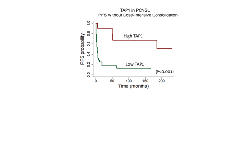
Using a variety of syngeneic and non-syngeneic preclinical models, including patient-derived CNS lymphoma cells, as well as diagnostic clinical specimens, we characterized the phenotype of tumor-associated macrophages in PCNSL. Using flow-cytometry, we demonstrated that while CD45 high tumor-associated macrophages exhibit strong expression of the canonical M2 marker CD206, a scavenger receptor, these also displayed high co-expression of iNOS and MHC II, markers of classically-activated M1 macrophages. Pharmacologic inhibition of the CSF-1 receptor led to accelerated CNS lymphoma progression, attenuated T-cell infiltration and blocked rituximab efficacy. A flow-cytometric assay of phagocytosis, using Raji lymphoma transduced to express mCherry, demonstrated that infiltrating CD206+ macrophages are the dominant mediator of lymphoma phagocytosis. We applied 2P intravital imaging of a CNS lymphoma model using Cx3cr1GFP/+:Ccr2RFP/+ myeloid cell dual reporter mice and transcriptional studies to define the time-dependent infiltration and phenotypic changes in tumor-associated macrophages and microglia that correlate with disease progression. Using IFN-γ -/- mice we identified a critical role for IFN-γ in the regulation of CNS lymphoma, in the presence and absence of T-cells. We identified IFN-γ-regulated genes in tumor-associated macrophages that may contribute to direct lymphoma cytotoxicity as well as stimulation of T-cell chemotaxis and antigen processing, including TAP1 and TAP2. By IHC, we confirmed TAP1 expression in a subset of diagnostic specimens of PCNSL and determined, using Cox multivariate model, that strong TAP1 correlated with improved PFS (p<0.0006). Notably, independent of receipt of maintenance therapy, TAP1 also correlated with improved PFS in 38 patients that received only MTX-based induction, without dose-intensive chemotherapy consolidation. (Figure 1)
Our results support a direct, immune-editing role for monocyte-derived macrophages in the regulation of CNS lymphoma progression, via several mechanisms, including antigenic processing and cross-presentation. We suggest that tumor-associated CD68 and TAP1 (and TAP2) be evaluated further as candidate biomarkers for risk stratification in PCNSL, particularly in trials that involve targeted immunotherapy. Supported by NCI and LLS.
Rubenstein: Kymera: Research Funding.


This feature is available to Subscribers Only
Sign In or Create an Account Close Modal